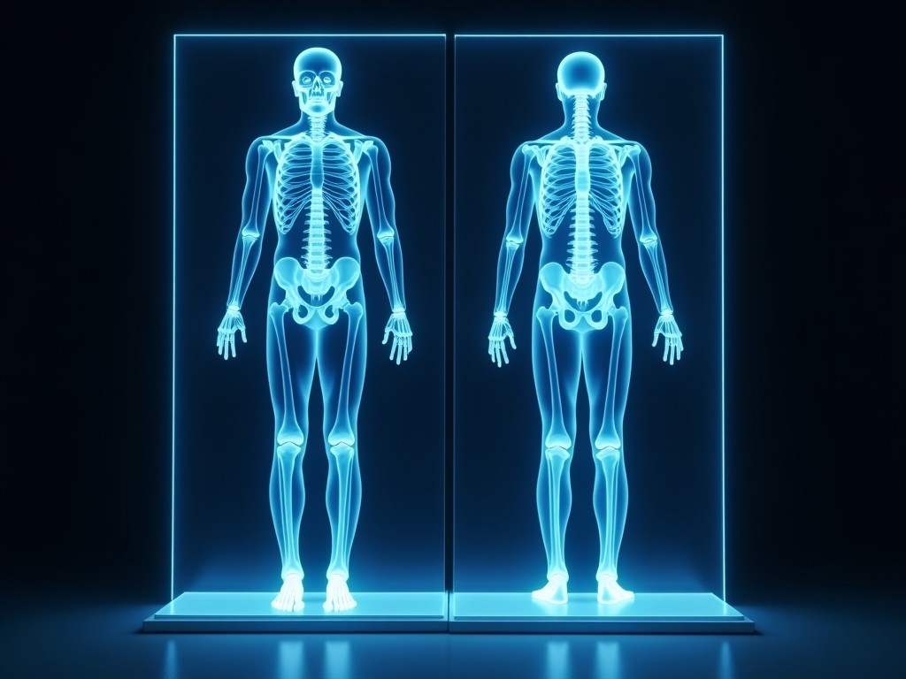
Oem 3D Body Scan Vs Dexa
Both 3D body scanning and dual‑energy X‑ray absorptiometry (DEXA) are non‑contact methods used to assess body composition, but they differ in measurement principle, spatial resolution, and typical
Inquire NowOEM 3D Body Scan vs. DEXA: Technical Comparison for Industrial Applications
Overview
Both 3D body scanning and dual‑energy X‑ray absorptiometry (DEXA) are non‑contact methods used to assess body composition, but they differ in measurement principle, spatial resolution, and typical industrial use cases. Understanding these differences helps OEMs select the technology that aligns with product development goals, regulatory constraints, and downstream data processing requirements.
A 3D body scan captures surface geometry using structured light, stereoscopic vision, or time‑of‑flight sensors, producing a point cloud or mesh that represents external shape. DEXA, by contrast, emits low‑dose X‑ray beams at two energy levels to quantify bone mineral density and soft‑tissue composition through attenuation measurements.
Technology Principles
Structured‑light scanners project a known pattern onto the subject and analyze deformation via cameras, enabling sub‑millimeter surface detail without ionizing radiation. The acquisition time is typically under one second per frame, allowing dynamic capture of movement.
DEXA relies on the differential attenuation of high‑ and low‑energy photons by bone and soft tissue. The system calculates bone mineral content (BMC) and lean/fat mass ratios based on attenuation coefficients, providing a volumetric estimate of internal composition.
Because 3D scanning measures only the exterior surface, it cannot directly assess internal tissue density, whereas DEXA provides internal compositional data but lacks surface shape detail beyond approximate contours.
Measurement Accuracy and Precision
Typical 3D scanners achieve point‑to‑point repeatability of 0.1 mm to 0.5 mm depending on sensor baseline and lighting conditions. Surface texture and reflectivity can introduce noise, requiring post‑processing filters or multi‑view fusion.
DEXA precision for bone mineral density is commonly expressed as a coefficient of variation (CV) of 1 % to 2 % for lumbar spine measurements, with soft‑tissue mass estimates showing CVs of 2 % to 4 % under standardized positioning.
When comparing volumetric fat mass, 3D scanning derives estimates from surface‑based models (e.g., volume × assumed density) leading to larger prediction errors (±5 % to ±10 %) compared with DEXA’s direct attenuation‑based quantification (±2 % to ±3 %).
For applications requiring external morphology (e.g., ergonomic fitting, garment sizing), 3D scanning’s geometric fidelity is advantageous. For dosing calculations based on bone strength or metabolic risk, DEXA’s internal compositional accuracy is preferable.
Application Suitability
Industrial sectors that benefit from high‑resolution surface data include aerospace seat design, protective equipment manufacturing, and custom orthotics, where the external shape drives fit and comfort.
DEXA finds use in pharmaceutical clinical trials, nutritional product development, and occupational health monitoring where bone health or fat‑lean ratios are critical endpoints.
Some OEMs combine both modalities: a quick 3D scan for size classification followed by a targeted DEXA scan for subjects falling outside predefined thresholds, optimizing throughput while retaining compositional insight.
Specification Comparison Table
| Parameter | OEM 3D Body Scan | DEXA |
|---|---|---|
| Measurement Principle | Structured light / stereo vision / ToF | Dual‑energy X‑ray attenuation |
| Primary Output | Point cloud / mesh (surface geometry) | BMD, BMC, lean mass, fat mass |
| Spatial Resolution (surface) | 0.1 mm – 0.5 mm | Not applicable (internal) |
| Depth Penetration | Surface only (≤ few mm depending on penetration of light) | Whole body (bone and soft tissue) |
| Radiation Dose | None | Low dose (≈ 1 µSv per scan) |
| Typical Scan Time | < 1 s per frame | 5 – 15 s (including positioning) |
| Repeatability (CV) | 0.1 mm – 0.5 mm absolute | 1 % – 2 % (BMD) |
| Typical Use Case | Ergonomic fitting, custom apparel, VR avatars | Bone health assessment, body composition trials |
Integration and Customization
OEM 3D scanning systems are offered with SDKs that provide raw point clouds, mesh generation, and alignment APIs, enabling integration into robotic cells or quality‑control stations. Lens options, lighting modules, and calibration targets can be adapted to specific part sizes or environmental constraints.
DEXA units typically come with calibrated phantoms and software packages for whole‑body analysis. Customization focuses on positioning aids, patient‑specific protocols, and export formats (DICOM, CSV) for downstream statistical packages.
Both platforms support firmware updates and can be housed in enclosures meeting IP ratings appropriate for industrial environments (e.g., IP54 for dust‑resistant scanning booths).
FAQ
Can the 3D scanner be used for volumetric fat estimation?
Yes, by applying assumed tissue density to the segmented volume, but accuracy depends on hydration status and posture; validation against DEXA is recommended for critical applications.
Is DEXA safe for repeated use in occupational health monitoring?
The effective dose per scan is low (≈1 µSv), allowing periodic monitoring within regulatory limits, though cumulative exposure should be tracked according to local guidelines.
What file formats are available for scan data?
Typical outputs include STL, OBJ, PLY for meshes and XYZ or CSV for point clouds; custom formats can be arranged per project requirements.
Contact for Inquiry
To discuss how either technology can be integrated into your product line, share your application requirements, part dimensions, and desired throughput. Our engineering team will evaluate feasibility, suggest optimal configuration, and provide a quotation.
Please use the contact form or email us at [email protected] to begin the conversation.