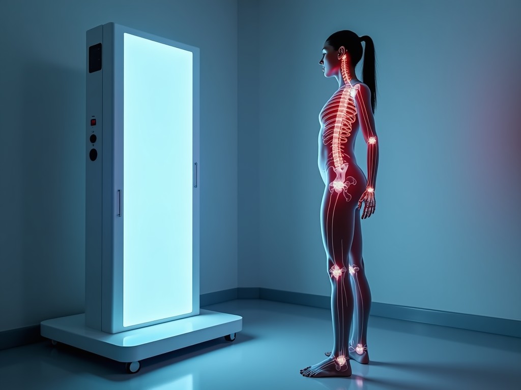
Dexa Body Composition Analysis Machine
A dual‑energy X‑ray absorptiometry (DEXA) system measures bone mineral density and soft‑tissue composition by exposing the subject to two narrow‑energy X‑ray peaks. The differential attenuation of
Inquire NowDEXA Body Composition Analysis Machine
Overview
A dual‑energy X‑ray absorptiometry (DEXA) system measures bone mineral density and soft‑tissue composition by exposing the subject to two narrow‑energy X‑ray peaks. The differential attenuation of these peaks allows the system to separate bone, lean tissue, and fat mass with a spatial resolution typically better than 5 mm.
Core Technology
The scanner employs a fan‑beam X‑ray tube operating at 80 kVP and 100 kVP, switching rapidly between the two energies. A linear detector array captures the transmitted photons, and proprietary algorithms compute pixel‑wise material fractions. The system incorporates a temperature‑stabilized reference detector to correct for drift, ensuring long‑term stability within ±0.5 % over 12 months.
Key Technical Specifications
| Parameter | Typical Value | Notes |
|---|---|---|
| Scan Area (L × W) | 200 mm × 800 mm | Covers whole‑body adult subjects |
| Spatial Resolution | ≤5 mm (FWHM) | Determined by detector pitch and focal spot size |
| Precision (CV%) | ≤1 % for lean mass, ≤1.5 % for fat mass | Based on repeat scans of phantom |
| Radiation Dose per Scan | 1–3 µSv (equivalent to < 0.1 mrem) | Well below annual occupational limits |
| Scan Time | 4–7 s for whole body | Includes positioning and acquisition |
| Power Requirements | 100–240 V AC, 50/60 Hz, 300 VA | Single‑phase, IEC 60320 C13 inlet |
| Dimensions (W × D × H) | 800 mm × 600 mm × 1800 mm | Includes detector gantry and console |
| Weight | 180 kg | Floor‑standing, requires leveling to ±2 mm |
Typical Industrial Applications
In pharmaceutical research, DEXA provides non‑invasive quantification of muscle wasting or adiposity in preclinical studies, enabling dose‑response modeling without sacrificing animals. In occupational health programs, the machine tracks changes in lean mass among workers undergoing physical‑training interventions, offering objective data for ergonomic assessments. Sports‑science laboratories use the precise regional‑fat‑distribution output to evaluate training periodization effects on specific muscle groups.
Measurement Precision and Repeatability
The system’s coefficient of variation for repeated measurements on a tissue‑equivalent phantom is typically below 1 % for lean mass and 1.5 % for fat mass when scans are performed under identical positioning conditions. This level of repeatability supports longitudinal studies where a change of 2 % in body composition is considered biologically significant. The built‑in auto‑positioning laser guides reduce operator‑induced variance to less than 0.3 %.
Software and Data Output
The acquisition workstation runs a Windows‑based platform that exports results in DICOM, CSV, and PDF formats. A built‑in XML‑compatible API allows integration with laboratory information management systems (LIES) for automated batch processing. The software provides voxel‑level maps of bone mineral density (BMD), lean tissue index (LTI), and fat tissue index (FTI), with optional sub‑region analysis (e.g., android vs. gynoid fat).
Installation and Site Requirements
A dedicated room of at least 3 m × 3 m with a flat, vibration‑isolated floor is recommended. The scanner requires a minimum ceiling height of 2.4 m to accommodate the gantry’s vertical travel. Shielding is not mandatory for the low dose levels, but a lead‑lined wall (0.5 mm Pb equivalent) may be installed if local regulations demand it. Ambient temperature should be maintained between 18 °C and 24 °C, with relative humidity below 60 % to prevent condensation on optical components.
Quality Assurance and Calibration
Each unit undergoes a factory acceptance test that includes a uniformity check across the detector array using a calibrated aluminum step wedge. Field calibration is performed quarterly with a NIST‑traceable phantom containing known concentrations of bone, lean, and fat equivalents. The calibration routine adjusts gain offsets and energy‑response coefficients, maintaining accuracy within ±0.02 g/cm² for BMD measurements.
Customization Options
- Optional whole‑body vibration platform for dynamic loading studies
- Extended software module for visceral adipose tissue (VAT) estimation
- Wireless handheld remote for patient positioning assistance
- Customizable tabletop finish (powder‑coated RAL colors) to match facility branding
- Extended warranty and on‑site preventive maintenance plans
Frequently Asked Questions
Can the scan area be increased for larger subjects?
The standard gantry accommodates subjects up to 200 mm in width; extensions are available upon request, increasing the width to 250 mm with a corresponding adjustment to the detector panel and software calibration.
What information is required before quotation?
To generate a accurate quote we need the intended scan throughput (scans per day), any required ancillary equipment (e.g., vibration platform, remote), and facility specifics such as power availability and room dimensions.
How is quality inspected?
Quality inspection includes visual examination of mechanical tolerances, functional verification of X‑ray output uniformity, and software validation against a multi‑material phantom. All test records are retained for the equipment’s lifecycle.
Next Steps
Engineers and procurement managers who require a reliable, low‑dose method for quantifying body composition can request a detailed technical datasheet or arrange a live demonstration. Please use the contact form below to initiate a discussion about configuration, lead time, and site‑specific adaptations.
Request Technical Datasheet