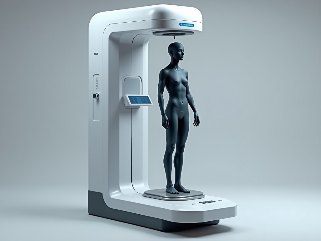
3D Body Composition Scanner Machine
The device captures a full‑body point cloud using structured light projection while simultaneously measuring multi‑frequency bioelectrical impedance across segmental electrodes. This hybrid approach
Inquire Now3D Body Composition Scanner Machine
The device captures a full‑body point cloud using structured light projection while simultaneously measuring multi‑frequency bioelectrical impedance across segmental electrodes. This hybrid approach yields voxel‑level tissue maps that are converted into fat mass, lean mass, total body water, and bone mineral estimates with segmental resolution.
Technology Overview
Structured light grids are projected onto the subject and captured by dual‑camera stereo sensors at 30 frames per second, producing a depth map with sub‑millimeter precision. Concurrently, eight surface electrodes inject alternating currents from 1 kHz to 1 MHz; the resulting voltage phases are used to compute intracellular and extracellular fluid volumes for each limb and torso segment.
Fusion of geometric and impedance data occurs in a calibrated algorithm that corrects for posture‑induced shadowing and electrode contact variance, delivering repeatability within ±1.5% for total body fat percentage across repeated scans.
Scan duration is limited to 12 seconds per subject, minimizing motion artifacts while accommodating high‑throughput environments such as sports clinics or wellness centers.
Key Specifications
| Parameter | Typical Value | Unit | Notes |
|---|---|---|---|
| Scan Volume | 2.0 m × 0.9 m × 0.6 m | m | Adjustable via optional extension frames |
| Depth Resolution | 0.3 | mm | At 1 m distance, RMS error |
| Impedance Frequencies | 1, 5, 50, 250, 500, 1000 | kHz | Six frequencies for segmental analysis |
| Measurement Repeatability (Fat %) | ±1.5 | % | Test‑retest on same day, same subject |
| Power Consumption | 120 | W | Average during scan |
| Operating Temperature | 10‑35 | °C | Non‑condensing humidity 20‑80% |
| Device Weight | 45 | kg | Includes base and sensor bar |
Applications
In sports performance labs the scanner provides segmental lean mass asymmetry data that correlates with injury risk, allowing trainers to adjust load distribution before maladaptive patterns develop.
Clinical nutrition programs use the visceral fat estimate derived from trunk impedance to monitor metabolic syndrome progression without ionizing radiation.
Ergonomics researchers employ the full‑body shape capture to create digital manikins for workspace design, ensuring that reach envelopes accommodate the 5th to 95th percentile of the target population.
Academic studies in human biology leverage the repeatable water compartment split to validate hydration protocols across varying environmental stressors.
Measurement Process
Prior to scanning, subjects remove outer clothing and stand on the calibrated footplate with heels aligned to the rear markers; this ensures consistent electrode contact and pose reproducibility.
The operator initiates a scan via the touchscreen console; structured light stripes sweep the volume while a synchronized current pulse trains through the electrode array, capturing both geometry and impedance streams in real time.
Raw point clouds undergo noise filtering and surface reconstruction; impedance data are segmentally integrated with the corresponding volume elements to compute tissue‑specific conductivity values.
Final outputs include regional fat‑free mass, fat mass, extracellular water, intracellular water, and a 3D avatar that can be exported in OBJ or STL formats for further analysis.
Customization Options
- Extended scan volume up to 2.5 m height for accommodating basketball players or specialized gait analysis setups.
- Wireless electrode modules enabling scapular or cranial impedance measurements for research protocols.
- Software packages offering API access for HL7 or FHIR integration with electronic health record systems.
- Enclosure materials: powder‑coated steel for industrial settings or antimicrobial polymer for clinical environments.
All options are configurable at quote stage; lead times vary from two to six weeks depending on complexity.
Quality Control & Calibration
Each unit undergoes a three‑point length verification using a calibrated gauge block set before leaving the factory, confirming depth accuracy within ±0.2 mm across the field of view.
Impedance channels are checked against precision resistor networks traceable to national standards, ensuring phase error below 0.5 degrees at all test frequencies.
Firmware includes a self‑diagnostic routine that runs at startup, reporting sensor health, laser power stability, and electrode contact impedance; any deviation beyond thresholds triggers a service alert.
Recommended field verification interval is 12 months, using a phantom with known tissue‑equivalent properties to validate both geometric and bioimpedance measurement chains.
Frequently Asked Questions
Yes, optional extension rails increase the maximum height to 2.5 m without compromising depth resolution.
OBJ, STL, and PLY formats are standard; additional formats such as VRML can be supplied upon request.
Installation, calibration, and operator training are included in the purchase package; scheduling is coordinated after order confirmation.