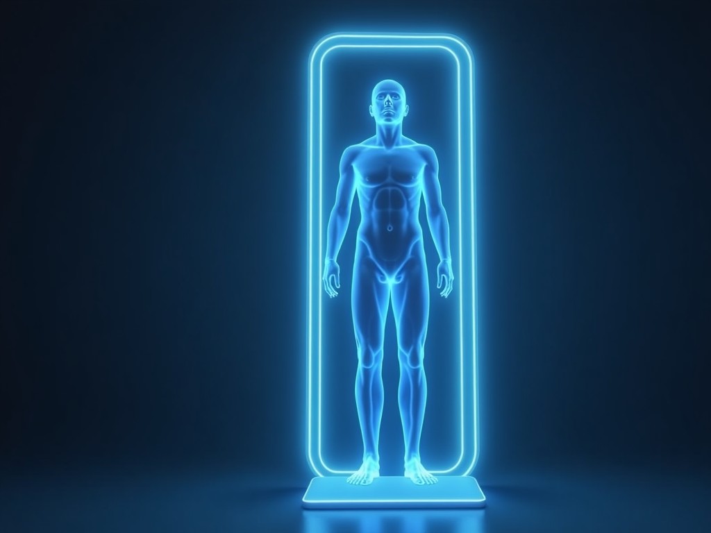
Wholesale 3D Body Fat Scanner
A wholesale 3D body fat scanner combines structured‑light depth imaging with multi‑frequency bioelectrical impedance to capture both external body volume and internal tissue conductivity in a single,
Inquire NowWholesale 3D Body Fat Scanner
A wholesale 3D body fat scanner combines structured‑light depth imaging with multi‑frequency bioelectrical impedance to capture both external body volume and internal tissue conductivity in a single, non‑contact scan. The resulting data set enables estimation of total body fat percentage, lean mass, and segmental composition without the need for dyes, radiation, or physical contact.
Technical Overview
The scanner projects a patterned infrared grid onto the subject while a depth camera records the deformation of the pattern at up to 120 frames per second, yielding a voxel‑based mesh with a typical resolution of 2 mm. Simultaneously, an array of eight surface electrodes injects a low‑amplitude alternating current (5 kHz–500 kHz) and measures the resulting voltage to derive segmental impedance. By correlating the volumetric mesh with impedance spectra, the device calculates fat and lean tissue distribution using a validated four‑component model.
Key Specifications
| Parameter | Typical Value | Notes |
|---|---|---|
| Scan Volume | 0.5 m × 0.5 m × 2.0 m | Accommodates standing adult subjects |
| Body Fat Accuracy | ±2 % (vs. DXA) | Mean absolute error across BMI 18–35 |
| Repeatability (CV) | ≤0.5 % | Three consecutive scans, same subject |
| Scan Time | <10 s | Includes volume capture and impedance sweep |
| Output Formats | CSV, JSON, HL7 FHIR | Configurable via API |
| Power Supply | 100‑240 V AC, 50/60 Hz | Auto‑ranging, 120 W max |
| Operating Temperature | 0 °C – 40 °C | Non‑condensing |
| Dimensions (W×D×H) | 600 mm × 400 mm × 1800 mm | Floor‑standing unit |
| Weight | ≈45 kg | Includes base stabilizers |
| Interface | USB 3.0, Gigabit Ethernet | Plug‑and‑play with host PC |
| Compliance | CE, FCC (available upon request) | ISO 13485 certification project‑specific |
Applications
Fitness operators use the scanner to track members’ progress over time, providing objective data that supports personalized training programs and improves retention. Clinical settings benefit from the device’s ability to monitor fluid shifts and sarcopenia in patients undergoing dialysis or chemotherapy, where frequent DXA exposure is undesirable. Research laboratories employ the high‑speed volumetric capture for biomechanical studies, correlating external shape with internal composition in运动 science protocols.
Customization Options
- Electrode configuration: 8‑channel standard; optional 16‑channel array for higher spatial resolution
- Enclosure material: powder‑coated steel (standard) or antimicrobial ABS for healthcare environments
- Mounting choices: floor‑standing base, wall‑mounted bracket, or portable cart with lockable casters
- Software integration: RESTful API, HL7 interface, or white‑label mobile app for end‑user data access
- Calibration kits: includes reference phantom and software tools for field verification
Quality Assurance Philosophy
Each unit undergoes a two‑stage verification: first, the depth camera subsystem is tested against a calibrated gauge block array to confirm voxel accuracy within 0.1 mm; second, the bioimpedance front‑end is checked using precision resistors to ensure impedance measurement error remains below 1 % across the frequency spectrum. Environmental testing includes temperature cycling (‑10 °C to 50 °C) and humidity exposure (10 %–90 % RH) to validate long‑term stability. Final functional validation is performed by comparing scanner outputs with dual‑energy X‑ray absorptiometry on a cohort of 30 subjects representing varied BMI and ethnicity.
Frequently Asked Questions
- Can the scan volume be adjusted for different populations? The optical field of view is fixed; however, optional wide‑angle lenses are available upon request to increase height capacity for taller individuals.
- What information is required before quotation? Please provide intended usage environment, anticipated daily throughput, and any required software interfaces so we can configure the appropriate electrode count and enclosure.
- Which materials are available for the housing? Standard is powder‑coated steel; antimicrobial ABS or stainless steel grades 304/316 can be supplied depending on hygiene requirements.
- How is quality inspected before shipment? Each unit receives a full functional test, a safety dielectric withstand test, and a visual inspection per IPC‑A‑610 Class 2 criteria.
- What is the typical production lead time? Lead time ranges from 4 to 6 weeks for standard configurations; customized orders may require an additional 2‑3 weeks for tooling and validation.
Next Steps
To receive a detailed quotation, discuss customization, or arrange a sample evaluation, please contact our sales team via the inquiry form. Provide your company name, contact person, and specific application details so we can tailor the proposal to your operational requirements.