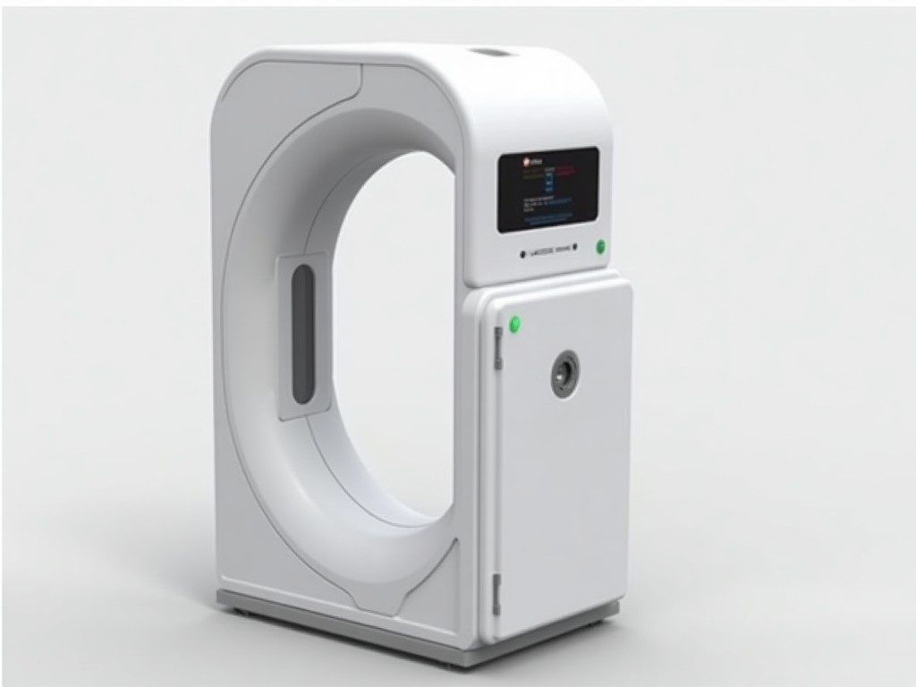
Buy Custom 3D Body Fat Scanner Machine
The device captures a dense point cloud of the human torso and limbs using structured light projection and multi‑camera triangulation. Each scan yields a volumetric model with sub‑millimeter surface
Inquire NowCustom 3D Body Fat Scanner Machine
The device captures a dense point cloud of the human torso and limbs using structured light projection and multi‑camera triangulation. Each scan yields a volumetric model with sub‑millimeter surface detail, from which tissue densities are inferred via calibrated empirical relationships.
Scanning Technology
The system employs a fringe‑pattern projector operating at 5 mm fringe pitch and two synchronized 120 fps infrared cameras. Point‑cloud density averages 8 000 points per cm², providing sufficient resolution to distinguish subcutaneous fat layers as thin as 2 mm.
Typical Measurement Output
From the reconstructed volume the scanner estimates total fat mass, lean mass, and segmental distribution (arms, legs, trunk). Repeatability tests show a coefficient of variation below 1.5 % for fat mass across successive scans taken within five minutes.
Technical Specifications
| Parameter | Typical Value | Notes |
|---|---|---|
| Scanning volume | 0.5 m × 0.5 m × 0.6 m (W×D×H) | Adjustable via lens change |
| Scan time | < 8 seconds per full body | Includes data transfer |
| Resolution (point spacing) | 0.3 mm | At nominal distance |
| Power consumption | 120 W (average) | 100‑240 V AC |
| Operating temperature | 10 °C – 35 °C | Non‑condensing |
| Data output | CSV, JSON, PLY via Ethernet | SDK available for integration |
Customization Options
- Alternative illumination wavelengths (visible or NIR) for specific skin‑type compensation
- Enlarged scanning envelope up to 0.8 m × 0.8 m × 1.0 m for full‑body or wheelchair access
- Integration of load‑cell platform for simultaneous weight measurement
- Enclosure material choices: powder‑coated steel or stainless‑steel for clean‑room environments
- Software modules: longitudinal tracking, clothing‑fit simulation, API for third‑party health platforms
Industrial Applications
Fitness facilities use the scanner to provide members with objective baseline data, reducing reliance on calipers or bio‑impedance devices that suffer from hydration variability. Apparel manufacturers leverage the detailed surface mesh to generate size‑specific patterns, decreasing sample‑making iterations by up to 30 %. In clinical research, the device offers a radiation‑free alternative to DXA for longitudinal adipose‑tissue studies, with scan‑to‑scan variance comparable to established methods.
Quality Assurance and Calibration
Each unit undergoes a factory verification using a certified phantom with known fat‑equivalent density. The verification checks volumetric accuracy within ±0.5 % and repeatability within ±0.2 % over ten consecutive scans. Field calibration kits, including a reference plate and software utility, are available for periodic user verification.
Frequently Asked Questions
Can the scanning volume be adjusted after purchase?
Yes, the lens system and camera positioning are modular; a service technician can reconfigure the envelope to meet new spatial constraints.
What file formats are available for raw data?
The scanner exports the point cloud as PLY or OBJ, and the processed composition report as CSV or JSON. Custom formats can be generated via the provided SDK.
Is training required to operate the device?
A standard operating procedure takes less than 30 minutes to learn. The interface guides the user through pose alignment, scan initiation, and result review.
To discuss your specific configuration or request a detailed quotation, please contact our sales team.
Request a Quote