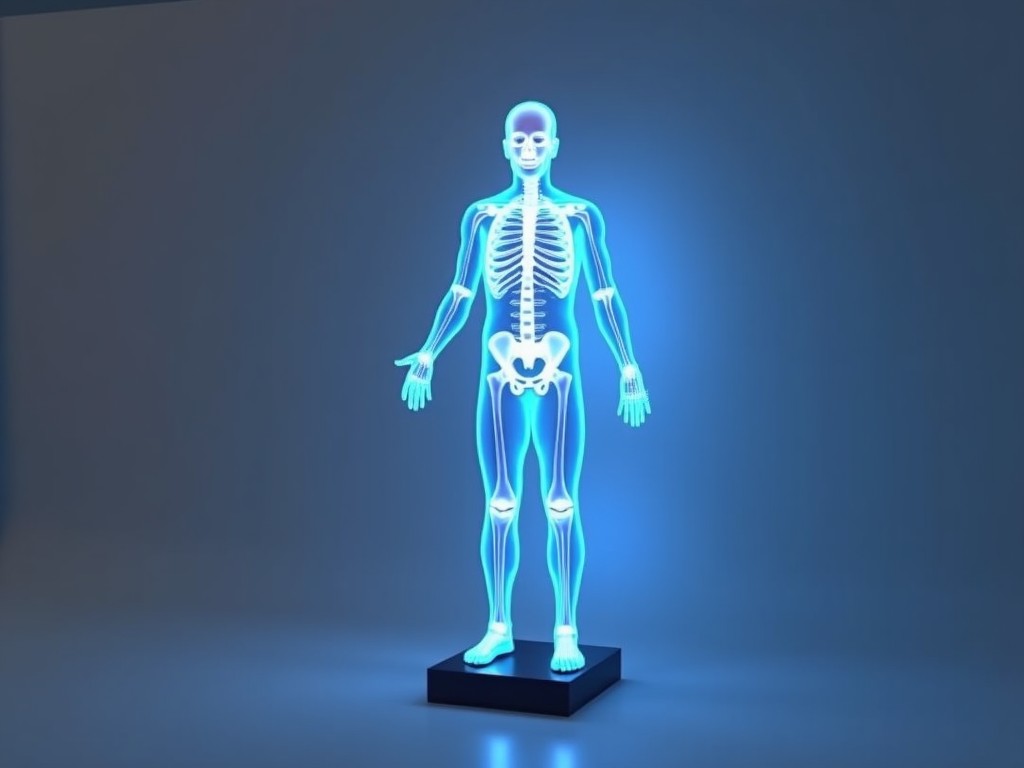
Buy 3D Body Fat Scanner Machine
The device captures a three‑dimensional surface map of the body while simultaneously applying multi‑frequency bioelectrical impedance analysis (BIA). By correlating volume data from the 3D scan with
Inquire Now3D Body Fat Scanner Machine
The device captures a three‑dimensional surface map of the body while simultaneously applying multi‑frequency bioelectrical impedance analysis (BIA). By correlating volume data from the 3D scan with impedance measurements at several frequencies, it estimates total body water, extracellular water, and fat mass with segmental resolution. This combined approach reduces reliance on empirical population‑based equations and improves accuracy across diverse body types.
Measurement Principle
A structured‑light projector and dual cameras generate a dense point cloud of the subject’s surface in under one second. Eight electrodes placed on the hands and feet inject alternating currents ranging from 5 kHz to 500 kHz; the resulting voltage drop is measured to calculate segmental impedance. The scanner’s software fuses the geometric volume and impedance data using a calibrated biophysical model to output fat‑free mass, fat mass, and visceral fat rating.
Key Specifications
| Parameter | Typical Value |
|---|---|
| Scan time | 0.8 s (surface) + 0.2 s (impedance) |
| Volume measurement range | 0.3 m³ to 0.15 m³ (≈30 kg to 150 kg mass) |
| Impedance frequencies | 5, 50, 100, 200, 500 kHz (5‑point) |
| Segmental resolution | 5 segments (trunk, arms, legs) |
| Repeatability (CV) | ≤1.5 % for fat mass |
| Power consumption | 15 VA max |
| Operating temperature | 10 °C – 35 °C |
| Interface | USB 2.0, Ethernet (optional) |
| Software compatibility | Windows 10/11, SDK available for custom integration |
Typical Applications
Fitness centers use the scanner to track members’ body composition changes over training cycles, providing objective data that supersedes skinfold calipers. In clinical settings, the device assists in monitoring edema, sarcopenia, and metabolic syndrome by delivering segmental fluid distribution. Research laboratories benefit from the high repeatability when studying the effects of nutrition or pharmaceutical interventions on adipose tissue.
Customization Options
- Electrode layout – standard hand‑to‑foot or optional hand‑to‑hand for upper‑body focus.
- Scanning area – adjustable field‑of‑view to accommodate seated or supine positions.
- Software modules – basic report, longitudinal tracking API, or HL7 interface for hospital information systems.
- Enclosure materials – powder‑coated steel for gym environments or antimicrobial ABS for clinical use.
Quality Assurance
Each unit undergoes a two‑point impedance verification using precision resistors traceable to national standards. The optical subsystem is checked with a calibrated flat‑field target to ensure point‑cloud density variation stays below 2 % across the scan volume. Firmware includes a self‑diagnostic routine that logs electrode contact quality and alerts the operator if impedance exceeds predefined thresholds.
Frequently Asked Questions
Can the scanning volume be adjusted for pediatric subjects?
Yes, the field‑of‑view can be reduced via software scaling, maintaining accuracy down to approximately 0.05 m³.
What is required before a quotation is issued?
Please provide the intended application, electrode configuration preference, and any required software interfaces.
How is the device shipped to prevent damage?
Units are packed in double‑wall corrugated boxes with interior foam inserts; each box includes a shock‑indicator label.