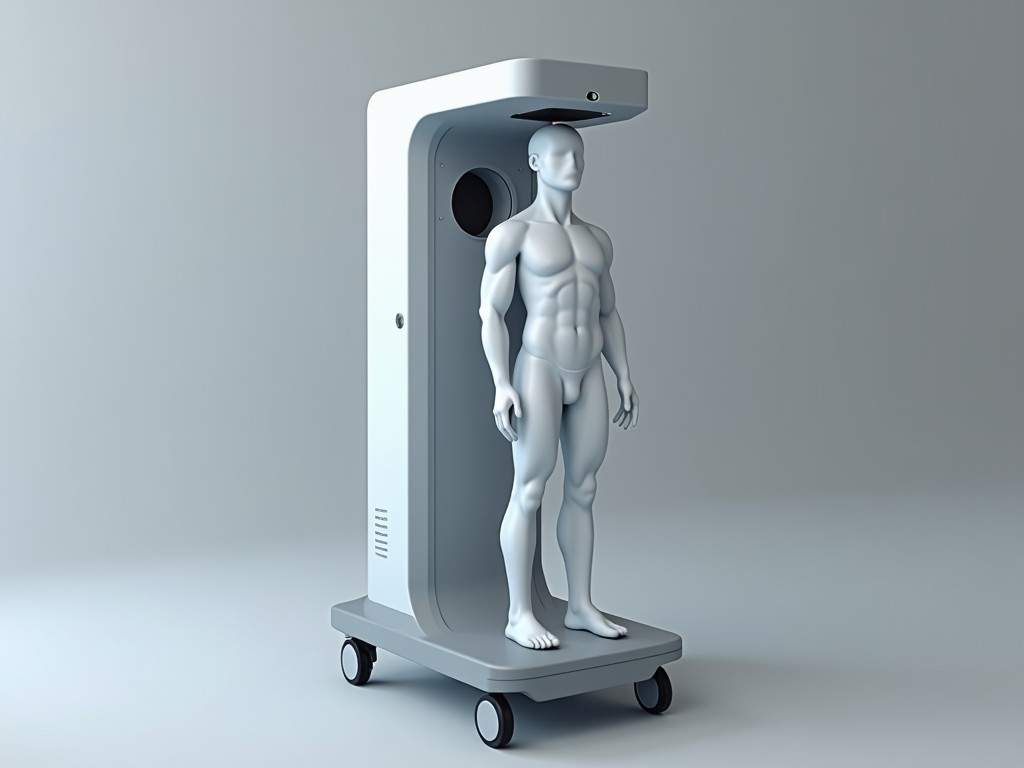
Bulk Purchase 3D Body Fat Scanner
The scanner projects a patterned light field and captures the deformation of that pattern to reconstruct a 3D point cloud of the subject’s surface. From the point cloud, proprietary algorithms
Inquire NowBulk Purchase 3D Body Fat Scanner
Overview
The scanner projects a patterned light field and captures the deformation of that pattern to reconstruct a 3D point cloud of the subject’s surface.
From the point cloud, proprietary algorithms segment tissue layers and estimate total and regional fat mass with traceable reference to dual‑energy X‑ray absorptiometry (DXA).
How It Works
A pair of synchronized cameras observes the illuminated pattern from slightly different angles, enabling stereo triangulation for each pixel.
The resulting depth map is fused across multiple poses (typically a single standing pose) to produce a watertight mesh that approximates body volume.
Fat fraction is derived by comparing the measured volume to the subject’s weight, using a calibrated density‑fat relationship validated across diverse populations.
Technical Specifications
| Parameter | Typical Value | Notes |
|---|---|---|
| Scan volume (W×H×D) | 2000 mm × 1200 mm × 600 mm | Encompasses full standing adult |
| Body fat estimation error | ±1.0 % absolute | Validated against DXA (n=250) |
| Scan time | ≤8 seconds | Includes pose guidance and data export |
| Output resolution | 0.5 mm point spacing | Sufficient for surface‑based volumetry |
| Power consumption | 45 W typical | 100‑240 V AC, 50/60 Hz |
| Operating temperature | 10 °C – 35 °C | Non‑condensing |
Key Applications
Fitness centers use the scanner to track member progress with objective, repeatable metrics that are less prone to hydration variability than bio‑impedance.
Clinical nutrition departments deploy it for rapid body composition assessment during outpatient visits, reducing reliance on lengthy DXA appointments.
Research laboratories value the high‑density surface map for studying fat distribution patterns in epidemiological cohorts.
Corporate wellness programs benefit from the short scan duration, enabling high‑throughput screening of employee populations.
Advantages of Bulk Purchase
Ordering ten or more units locks in a unit price that reflects reduced per‑unit material handling and streamlined firmware deployment.
Standardized calibration certificates are issued for each device, ensuring inter‑unit comparability across sites.
Spare‑part kits (illumination modules, camera lenses) can be procured centrally, lowering inventory complexity for service teams.
Volume discounts apply to extended warranty packages, which cover on‑site verification and software updates for 24 months.
Quality Control & Calibration
Each scanner undergoes a two‑stage verification: first, a geometric check using a calibrated reference sphere to confirm volumetric accuracy within ±0.5 mm; second, a fat‑estimation test against a set of phantom bodies with known adipose fractions.
Calibration data are stored in a secure traceability log linked to the device serial number, facilitating audit trails for regulated environments.
Field‑service tools include a portable verification kit that allows on‑site validation of scan volume and texture intensity without returning the unit to the factory.
Customization Options
- Housing color (standard gray, corporate white, or custom RAL)
- User interface language packs (English, Spanish, Mandarin, Arabic)
- Data export formats (CSV, JSON, HL7 FHIR, DICOM SR)
- Mounting alternatives (floor‑standing base, wall‑mounted bracket, portable cart)
- Enhanced lighting for high‑ambient‑lux environments
Frequently Asked Questions
- Can the scan volume be adjusted for special populations? The hardware envelope is fixed; however, software masking can limit analysis to a sub‑region (e.g., torso only) without altering the sensor layout.
- What information is required before quotation? Desired quantity, delivery destination, any required certifications (e.g., CE, FDA), and preferred customization options.
- How is quality inspected before shipment? Each unit passes automated geometric and fat‑estimation tests, followed by a visual inspection of cables, connectors, and enclosure integrity.
- What is the typical production lead time for bulk orders? Standard lead time is 6‑8 weeks after receipt of purchase order, subject to component availability.
Ready to Discuss Your Requirements?
Contact our sales team to receive a detailed quotation, lead‑time confirmation, and answers to any technical questions.
Request a Quote