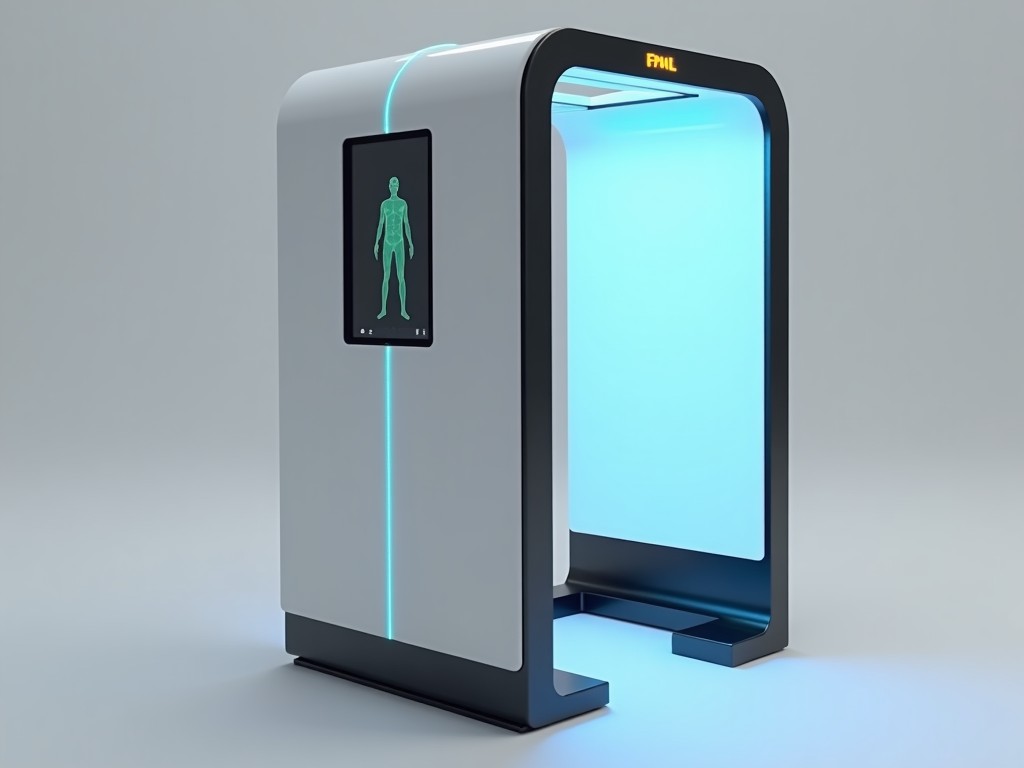
Budget Friendly 3D Body Scanning Mirror Machine
This device captures a full‑body point cloud by projecting a known pattern onto the subject and recording the deformation with two angled mirrors that feed a single‑camera system. The mirror
Inquire NowBudget Friendly 3D Body Scanning Mirror Machine
This device captures a full‑body point cloud by projecting a known pattern onto the subject and recording the deformation with two angled mirrors that feed a single‑camera system. The mirror arrangement doubles the effective baseline without increasing the physical footprint, allowing a compact unit to achieve metrology‑grade accuracy.
Operating Principle
A calibrated fringe pattern is emitted from a LED projector onto the person standing in front of the unit. Two front‑surface mirrors, positioned at 45°, redirect the reflected pattern to a single high‑resolution CMOS sensor. By analyzing phase shifts in the pattern, the system reconstructs depth information for each pixel, producing a dense 3D map of the body surface.
Technical Specifications
| Parameter | Typical Value | Notes |
|---|---|---|
| Scanning Volume | 2000 mm × 1000 mm × 600 mm | Adjustable via mirror tilt |
| Point Spacing | 0.5 mm | At nominal distance |
| Accuracy (ISO 10360‑2) | ±0.8 mm | After factory calibration |
| Capture Speed | < 2 s per full scan | Includes pattern projection and image acquisition |
| Light Source | LED projector, 650 nm, 10 W | Long‑life, low heat |
| Sensor Resolution | 2448 × 2048 px | Global shutter CMOS |
| Output Formats | OBJ, STL, PLY, XYZ | Selectable via software |
| Power Consumption | 45 W typical | 100‑240 V AC, 50/60 Hz |
| Dimensions (W × H × D) | 650 mm × 1300 mm × 300 mm | Includes base stand |
| Weight | 28 kg | Net weight |
Key Features
- Single‑camera design reduces cost and alignment complexity.
- Mirror‑based optical path doubles effective baseline without enlarging enclosure.
- Factory‑calibrated fringe projection yields sub‑millimeter accuracy.
- Rapid capture (<2 s) supports high‑throughput environments.
- Open‑format data output enables integration with CAD, ERP, or custom analytics pipelines.
- Low power draw and passive cooling allow continuous operation.
Typical Applications
In apparel manufacturing, the scanner provides precise body measurements for pattern making and virtual fitting, reducing reliance on manual tape measures and minimizing material waste. Fitness centers use the data to track longitudinal changes in body composition, offering members objective feedback beyond weight scales. Healthcare providers employ the captured geometry for prosthetic socket design, orthotic fitting, and post‑surgical monitoring, where repeatable, contact‑free surface data improves device comfort and clinical outcomes.
Customization Options
- Alternative mirror angles to adapt scanning volume for seated or supine subjects.
- Upgrade to higher‑resolution sensor for sub‑0.3 mm point spacing.
- Enclosure color and branding panels to match facility aesthetics.
- Software modules for automated measurement extraction (e.g., chest circumference, limb length).
- Integration kit for robotic arm mounting in automated production lines.
Quality and Reliability
Each unit undergoes a two‑stage verification: first, interferometric testing of the mirror assembly to confirm surface flatness within λ/10; second, a traceable artifact scan to validate volumetric accuracy against a calibrated gauge block array. The LED projector is selected for a minimum 50,000‑hour lumen maintenance, and the CMOS sensor features global shutter to eliminate motion artifacts during rapid capture cycles. Firmware includes built‑in diagnostics that flag illumination intensity drift or sensor temperature excursions, enabling predictive maintenance.
Frequently Asked Questions
Can the scanning volume be adjusted for different user heights?
Yes, the mirror tilt mechanism allows the effective field of view to be shifted vertically by ±150 mm without changing the unit’s footprint, accommodating users from 1400 mm to 2000 mm tall.
What file formats are available for downstream processing?
The scanner exports OBJ, STL, PLY, and plain XYZ point clouds. Formats are selectable via the companion software interface, and batch conversion scripts are provided for pipeline integration.
Is on‑site calibration required after installation?
The device ships with a factory‑traceable calibration certificate. A quick verification using the supplied reference plate takes less than five minutes and confirms that accuracy remains within ±0.8 mm; full recalibration is only needed after significant mechanical shock or lens replacement.