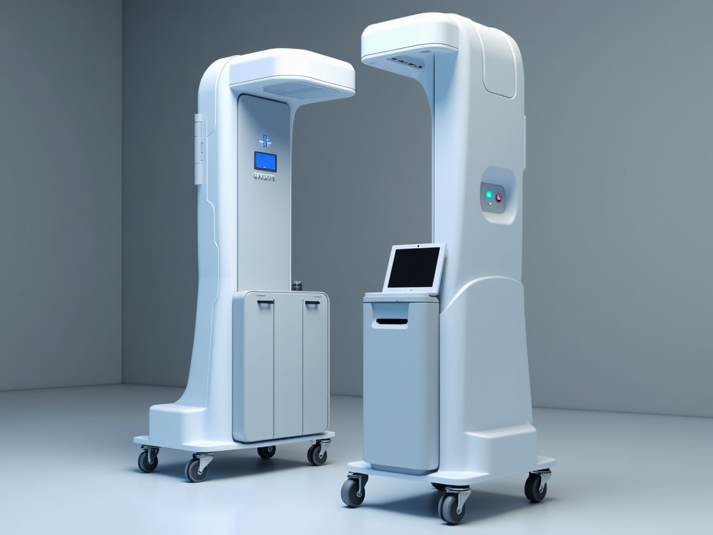
B2B 3D Body Scanner Medical Machine
A 3D body scanner captures the external geometry of a patient or subject using structured light or laser triangulation, producing a dense point cloud that can be processed into a watertight mesh. In
Inquire NowB2B 3D Body Scanner for Medical Applications
A 3D body scanner captures the external geometry of a patient or subject using structured light or laser triangulation, producing a dense point cloud that can be processed into a watertight mesh. In a B2B context, the device is sold to hospitals, research institutions, and medical device manufacturers who need accurate, repeatable surface data for ergonomic design, prosthetic fitting, or surgical planning.
Working Principle
The scanner projects a series of known patterns onto the subject; a pair of cameras records the deformation of these patterns from different viewpoints. By applying triangulation algorithms, the system calculates XYZ coordinates for each pixel, achieving sub‑millimeter spatial resolution. The process completes in under two seconds per scan, allowing dynamic studies such as breathing‑induced torso movement.
Key Technical Specifications
| Parameter | Typical Value | Notes |
|---|---|---|
| Scan Volume (W×H×D) | 500 mm × 500 mm × 500 mm | Adjustable via lens change |
| Spatial Resolution | 0.2 mm (point spacing) | At 1 m working distance |
| Accuracy (ISO 12836) | ±0.3 mm RMS | Verified on calibrated gauge block |
| Capture Speed | 1.5 s per full‑body scan | Includes data transfer |
| Light Source | Blue LED, 470 nm | Eye‑safe, low power |
| Output Formats | STL, OBJ, PLY, PTX | Binary or ASCII |
| Power Consumption | 45 W (typical) | 100‑240 V AC, 50/60 Hz |
| Operating Temperature | 10 °C – 35 °C | Non‑condensing |
| Weight (Scanner Unit) | 12 kg | Includes mounting bracket |
Clinical and Industrial Applications
In prosthetic and orthotic labs, the scanner provides a contact‑free capture of limb shape, reducing molding time and improving socket fit. Radiology departments use the data to create patient‑specific bolus markers for radiation therapy, ensuring consistent positioning across fractions. Ergonomics researchers employ repeated scans to quantify postural changes during rehabilitation exercises, enabling objective outcome measures.
Medical device manufacturers integrate the scanner into design verification pipelines, comparing virtual prototypes against actual user anatomy to assess clearance and comfort. The high repeatability (±0.1 mm between consecutive scans) supports statistical shape modeling for population‑based sizing studies.
Customization Options
- Lens kits for extended scan volume up to 800 mm cube
- Optional turntable with programmable rotation for full‑body automation
- Enclosure with antimicrobial coating for clinical environments
- Software development kit (SDK) for custom measurement plugins
- Calibration artifacts traceable to NIST standards
Quality Assurance and Calibration
Each unit undergoes a two‑stage verification: first, a geometric check using a calibrated step‑height gauge to confirm linearity across the measurement range; second, a repeatability test on a reference sphere to validate point‑to‑point stability. Calibration certificates are supplied with every shipment, and field recalibration is recommended every 12 months or after any mechanical shock.
The firmware includes built‑in diagnostics that monitor laser power, camera exposure, and temperature drift, issuing alerts via Ethernet if any parameter exceeds predefined thresholds. This proactive monitoring reduces unexpected downtime and helps maintain data integrity across longitudinal studies.
Integration and Software
The scanner ships with a Windows‑based control application that offers real‑time preview, alignment tools, and mesh‑cleaning filters (outlier removal, hole filling, smoothing). Data export is handled through a standardized API, allowing seamless connection to PACS, CAD platforms, or custom analytics pipelines. Network connectivity is via Gigabit Ethernet with optional PoE for simplified cabling.
Frequently Asked Questions
Can the scan volume be adjusted for pediatric patients? Yes, by swapping the lens kit and adjusting the working distance, the effective volume can be reduced to 250 mm³ while maintaining the same resolution.
What file formats are compatible with common CAD software? The scanner outputs STL and OBJ, which are directly importable into SolidWorks, AutoCAD, and Blender without conversion.
Is on‑site installation and training available? Installation and operator training can be arranged upon request; typical lead time is two weeks after purchase order confirmation.
How is data security handled for patient scans? All data transfer uses TLS‑encrypted channels; the control software stores files locally with optional automatic deletion after export.